Special Equipment
 Optomap®:
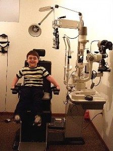
The Optomap® Retinal Exam provides a panoramic view of the back of a person’s eye (the retina). The view provided by the Optomap displays over 80% of the retina.
We have you place your eye to be photographed up to the instrument and the doctor’s assistant positions you for a photograph and takes the picture. The Optomap captures its image in only 1/4 of a second. The photographer takes two photos and evaluates the images. If the images are clear for the doctor to read the same thing is repeated on the second eye.
Click here for more information
 OCT:
OCT:
The OCT uses a method called “optical coherence tomography” that is capable of creating digital images through the use of special beams of light in order to create a contour map of the optic nerve and measure the retinal nerve fiber thickness much in the same way that a CT Scan is able to digitize and analyze tissues throughout your body.
The goal of Optic Nerve Computer Imaging is to give your eye the ability to detect the slightest loss of optic nerve fibers, at the first possible moment, in order to diagnose Glaucoma at the earliest possible stage in order to stop the progression of the disease and preserve your vision.
Visual Fields:
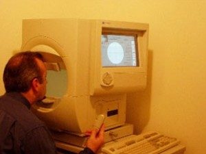
What is visual fields testing? For example, as you focus on the words in an article, how much can you see out of the corners of your eyes? Can you tell what’s happening in your surroundings? Your visual field is how wide of an area your eye can see when you focus on a central point.
Visual field testing is one way your optometrist measures how much vision you have in either eye and how much vision loss may have occurred over time. A visual field test can determine if you have blind spots (called scotoma) in your vision and where they are. A scotoma’s size and shape can show how eye disease or a brain disorder is affecting your vision. For example, if you have glaucoma, this test helps to show any possible side (peripheral) vision loss from this disease.
To do this test, you will look into the center of a bowl-shaped instrument called a perimeter. The eye not being tested will be covered with a patch. The testing eye will have your lens prescription placed in front of it to make sure you are seeing as well as possible.